Melanoma in Paris 8: screening, surgery and follow-up

Recognizing a suspicious spot, having it removed and analyzed, understanding surgical revision and follow-up: the complete process, explained by a plastic surgeon in Paris 8th arrondissement.

Table of contents

Contact us
at the clinic

Medically reviewed by Dr. Nathaniel Stroumza-Escoffier , plastic surgeon, RPPS 10100674943.

Published on May 15, 2025 · Updated on October 28, 2026.

Melanoma is a skin cancer that originates in the cells that produce pigment. It accounts for approximately one in ten skin cancers [ 2 ] . The thinner the tumor at the time of diagnosis, the better the prognosis [ 17 ] . Other skin cancers are described on the page dedicated to skin cancer.

Short answer

Melanoma cannot be definitively identified with the naked eye. A spot that changes, looks different from others, or bleeds should be shown to a doctor [ 2 , 4 ] . Diagnosis is based on the complete removal of the lesion and its analysis under a microscope , never on its destruction [ 15 , 16 ] . If melanoma is confirmed, a second procedure, called re-excision, removes a margin of healthy skin: 0,5 cm for melanoma in situ , 1 cm up to 1 mm thick, 1 to 2 cm between 1 and 2 mm, and 2 cm beyond that [ 2 , 18 ] . Follow-up of the skin then continues for life [ 2 ].

Key points

  • In France, 17,922 new cases of cutaneous melanoma and 1,904 deaths were estimated in 2023[1].
  • Net survival at 5 years reached 93%, across all stages, for people diagnosed between 2010 and 2015[1].
  • 7 to 8 melanomas out of 10 appear on healthy skin, and not on an existing mole[2, 3].
  • When examined with a dermatoscope by a trained physician, a spot is better classified: 92% of melanomas detected, compared to 76% to the naked eye, for the same false alarm rate[8].
  • A suspicious stain is never destroyed (laser, liquid nitrogen, cream): it is removed and analyzed.[15].
  • The thickness of the tumor, or Breslow index, determines the prognosis, the margin for recovery, and the indication for sentinel lymph node biopsy[2, 17, 20].

Definitions

Melanoma : skin cancer that develops from melanocytes, the cells that produce pigment [ 2 ].

Melanoma in situ : melanoma still limited to the epidermis, the superficial layer of the skin.

Breslow index : tumor thickness, measured under a microscope in millimeters. This is the main element of the prognosis [ 17 ].

Excision : surgical removal of the lesion. Re-excision is the second excision, which enlarges the removed area once the diagnosis is known [ 2 ].

Sentinel lymph node : the first lymph node to which the melanoma skin area drains. Its analysis provides information on the stage [ 20 ].

What is a melanoma?

Melanoma originates in melanocytes, the cells that give skin its color. It can metastasize to the lymph nodes or other organs [ 2 ] . Other skin cancers are carcinomas, which are much more common; cutaneous carcinoma is discussed on a separate page.

In France, the median age at diagnosis is 68 years for men and 62 years for women. The rate of new cases increased by 1,6% per year between 2019 and 2023 [ 1 ] . Melanoma can appear anywhere, including on areas with low sun exposure: palms, soles of the feet, nails [ 2 , 6 ].

The four main forms

The four main forms of melanoma
FormWhat there is to know
Superficial spreading melanomaThe most frequent form: from four out of ten cases to two out of three, according to the sources[2, 6].
Nodular melanomaRapid growth[2, 6]More often symmetrical, in relief, of a single color, sometimes with little pigmentation: the ABCDE rule poorly identifies it.[7].
Lentil melanomaEspecially on the face, in older people, linked to prolonged sun exposure[2, 6]Its profit margins follow a specific rule.
Acral lentiginous melanomaPalms, soles, nails, unrelated to the sun[2, 6]1 to 5% of melanomas occur on fair skin, with higher rates in Asian and African-American populations.[6].

Melanoma, mole or carcinoma?

Melanoma, mole, or carcinoma: the differences
Mole (nevus)MelanomaCarcinoma
NatureBenign melanocyte lesionMelanocyte cancerKeratinocyte cancer
Usual appearanceSymmetrical, homogeneous, stableChange, stands out from other spotsPinkish bump or scab that won't heal
Risk of metastasisNoReal, related to thicknessRare (basal cell), possible (squamous cell)
RacingMonitorExcision, analysis, re-excisionExcision, analysis

Only a medical examination, followed by microscopic analysis, can determine the cause [ 2 ].

What signs should prompt a consultation?

The ABCDE rule

The ABCDE rule summarizes the signs that should prompt a doctor to show a pigmented spot [ 2 , 4 ] :

  • A is for asymmetry : the two halves do not resemble each other;
  • B for irregular edges : cut-out outline, poorly defined;
  • C as in inhomogeneous color : several shades in the same stain;
  • D as in diameter : more than 6 mm;
  • E is for evolution : the spot changes in size, shape, color or texture, itches or bleeds.
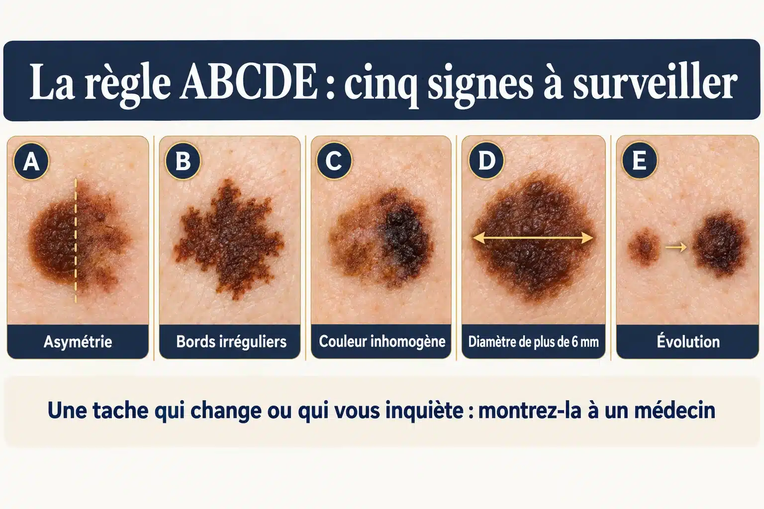
The ABCDE rule of melanoma illustrated by five skin lesions: asymmetry, irregular borders, uneven color, diameter greater than 6 mm, evolution
Figure 1 — The ABCDE rule: five signs that should prompt a doctor to examine a lesion. Diameter is a guideline, not a threshold; progression is the most important sign. Adapted from Abbasi et al. [4] and the National Cancer Institute [2]. Educational diagram.

Evolution is the most important sign. Diameter is a guideline, not a threshold: a melanoma often exceeds 6 mm, but not always [ 2 , 4 ] . Another indicator is the "ugly duckling": the spot that resembles no other in the same person [ 5 ].

What the ABCDE rule doesn't detect

Nodular melanoma is most often symmetrical, raised, and a single color [ 7 ] . For this type of melanoma, European experts propose the EFG rule: a raised, firm, enlarging lesion [ 6 ] . Some melanomas are not pigmented, which delays diagnosis [ 2 ] . These signs do not establish a diagnosis; they indicate that a consultation is necessary. Warning signs are detailed in the article on suspicious moles.

Consult without delay

Consult a doctor promptly, without waiting for a routine check-up, if a spot changes within a few weeks, if a raised, firm lesion grows larger, if a spot bleeds without injury, or if a new pigmented band appears on a nail. In the meantime, do not have the spot treated with laser, liquid nitrogen, or cream: it must be able to be analyzed [ 15 ].

Who is most at risk?

Ultraviolet radiation is the primary cause. In metropolitan France, 83,5% of melanomas in people over 30 were attributable to the sun in 2015, and 3,1% to tanning beds [ 29 ].

Risk factors for melanoma
SituationEstimated risk
101 to 120 moles (compared to fewer than 15)Approximately 7 times higher[9]
5 atypical moles (versus none)Approximately 6 times higher[9]
Personal history of melanomaApproximately 9 times more likely to have a second one[13] 2 to 4% of patients develop one[18]
Organ transplant, weakened immunityApproximately 2,7 times higher[12]
Close relative affectedApproximately 1,7 times higher[10] One in ten melanomas occurs in a family already affected by the disease.[2]
Tanning booth at least onceApproximately 20% higher[11]

Fair skin, freckles, red hair [ 10 ] , and severe sunburns in childhood [ 2 ] also increase the risk. These figures compare groups; they do not indicate the probability that a specific individual will develop melanoma.

For people at risk, the INCa recommends a self-examination of the skin every three months, i.e. about fifteen minutes, and an annual examination by a dermatologist or the attending physician [ 2 ].

Protect yourself from the sun

The INCa recommends avoiding the sun between noon and 4 p.m. in mainland France, seeking shade, and wearing protective clothing, a wide-brimmed hat, and sunglasses. On exposed areas, a sunscreen with SPF 50 should be reapplied every two hours. This complements other sun protection measures but does not replace them; tanning beds should be avoided [ 32 ].

How is the diagnosis made?

Dermatoscope and artificial intelligence

The doctor examines the spot with a dermatoscope, a magnifying glass with a light. When performed by a trained doctor, this examination detects more melanomas than the naked eye, without increasing the number of false alarms [ 8 ].

At the center, the examination is performed using the FotoFinder® digital dermatoscope, coupled with artificial intelligence to assist in analysis. Interpretation and decision-making remain the responsibility of medical professionals. The 2024 European consensus calls for caution regarding these tools [ 6 ] . In a Swiss study of 143 high-risk patients, the algorithm detected as many melanomas as dermatologists, but incorrectly classified approximately 35% of benign spots as suspicious, compared to 8% for dermatologists [ 14 ].

Diagnostic excision

Only microscopic analysis confirms the diagnosis. Therefore, when faced with a suspicious lesion, the physician removes it entirely from the outset, with a narrow 2 mm margin of healthy skin [ 2 ] . Partial removal remains the exception, for certain extensive lesions on the face or extremities [ 2 , 15 ] : it may underestimate the thickness of the tumor [ 15 , 16 ].

To remember

A spot whose suspicious nature cannot be ruled out cannot be destroyed: neither with a laser, nor with liquid nitrogen, nor with a cream [ 15 ] . A destroyed lesion can no longer be analyzed or measured. At the center, any removed lesion is sent to the laboratory.

The laboratory report specifies the thickness of the tumor and the presence or absence of an ulceration, a rupture of the epidermis visible under a microscope. These are the two elements used to classify it [ 17 ].

The Breslow thickness: why thickness matters so much

The Breslow thickness is the maximum thickness of the melanoma. It is measured under a microscope from the granular layer of the epidermis to the deepest tumor cell, rounded to the nearest tenth of a millimeter [ 2 , 17 ] . It is not measured for melanoma in situ , which has not penetrated beyond the epidermis [ 17 ].
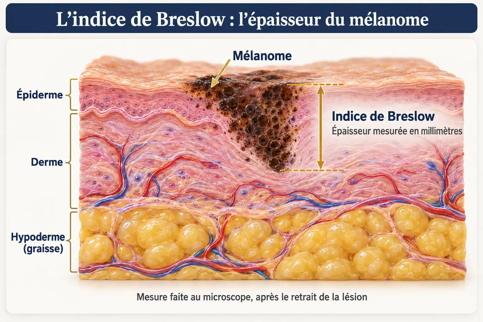
Cross-section of skin showing a melanoma extending from the epidermis into the dermis, with the vertical measurement of its thickness, the Breslow index.
Figure 2 — Breslow thickness: melanoma thickness, measured under a microscope from the granular layer of the epidermis (its upper part) to the deepest tumor cell. From the National Cancer Institute [2] and Gershenwald et al. [17]. Educational diagram.
Tumor thickness and category (AJCC international classification, 8th edition)
CategoryThickness (Breslow index)
On-siteLimited to the epidermis: no Breslow index
T1Up to 1 mm. T1a: less than 0,8 mm without ulceration. T1b: 0,8 to 1 mm, or less than 0,8 mm with ulceration.
T2More than 1 mm and up to 2 mm
T3More than 2 mm and up to 4 mm
T4More than 4 mm

International AJCC classification, 8th edition [ 17 ] . From T2 to T4, "a" means without ulceration and "b" means with ulceration.

The thinner the melanoma, the better the outcomes. In the international reference database, 99% of patients at 5 years and 98% at 10 years had not died from their stage IA melanoma. At stage IIC (larger than 4 mm, ulcerated, without lymph node involvement), these figures were 82% and 75%, respectively [ 17 ] . These figures come from specialized centers and, for tumors larger than 1 mm, from patients with unaffected sentinel lymph nodes, which makes them more favorable. They describe groups and do not predict the outcome for any individual.

The treatment: a two-stage surgery

The first step is to remove the lesion for analysis. This is the narrow-margin excision described above. It establishes the diagnosis and measures the thickness.

Second stage: revision. When melanoma is confirmed, revision is necessary in most cases, even if the first excision removed everything [ 2 ] . It removes a margin of healthy skin around the scar, which is held in place by the thickness of the melanoma.
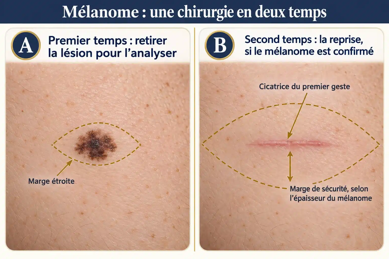
Melanoma surgery is performed in two stages: excision of the lesion with a narrow margin for analysis, followed by re-excision around the scar with a wider safety margin.
Figure 3 — Two-stage melanoma surgery: A, the lesion is removed with a narrow margin for analysis; B, if melanoma is confirmed, the second stage removes a margin of healthy skin around the scar, fixed according to the thickness of the tumor. From the National Cancer Institute [2]. Educational diagram.

The margins of recovery

Margins of re-excision depending on the thickness of the melanoma
Melanoma thicknessHealthy skin marginclarifications
On-site0,5 cmLentil melanoma in situ 1 cm; 0,5 cm accepted if anatomical requirements dictate, with strict histological control of the margins[2, 18]
Up to 1 mm1 cmEven for a very thin melanoma: 0,5 cm is only valid for thein situ[2, 18]
More than 1 mm and up to 2 mm1 to 2 cmEuropean guidelines recommend a 1 cm margin for any melanoma measuring 2 mm or less.[15, 19]
More than 2 mm2 cmA margin greater than 2 cm makes no difference.[2]

According to INCa (2024) [ 2 ] and the French Society of Dermatology (2016) [ 18 ] . Margins are measured on the skin, and not on the laboratory report.

In depth, the re-excision extends to the muscle sheath, without removing it. On the face and extremities, narrower margins may be necessary to preserve function; this adaptation is decided on a case-by-case basis [ 2 , 15 ] . Re-excision prevents recurrence in the same location; it does not affect the risk of distant metastasis [ 2 ].

Deadline and closing

French guidelines do not specify a numerical timeframe. The European consensus recommends performing reoperation preferably within 4 to 6 weeks of diagnosis, with sentinel lymph node biopsy when indicated [ 15 ] . Two observational studies showed no effect on survival of a delay of a few weeks before reoperation [ 30 ] or before sentinel lymph node biopsy [ 31 ].

Depending on the area and extent of skin loss, closure is achieved through direct suturing, flap surgery (using a nearby repositioned skin flap), or skin grafting. This falls under the umbrella of reconstructive surgery.

The sentinel lymph node: for whom?

The sentinel lymph node is the first lymph node to which the melanoma area drains. Removing it for analysis is not systematic: the indication depends on the thickness [ 2 , 20 ].

Sentinel lymph node detection based on thickness
SituationSentinel lymph node biopsy
Less than 0,8 mm, without ulceration (T1a)No indication[20]
0,8 to 1 mm, or less than 0,8 mm with ulceration (T1b)Can be offered[20]
More than 1 mm, without a palpable lymph nodeRecommended when additional treatment is being considered[20]

For melanomas 1 mm or smaller, the sentinel lymph node contained tumor cells in approximately 4,5% of cases [ 21 ] . Its analysis provides information on prognosis and the benefit of adjuvant treatment, but a large trial did not show that it improves overall patient survival [ 22 ] . If the sentinel lymph node is involved, other lymph nodes are no longer routinely removed [ 20 ] : this dissection did not improve survival and increased the risk of lymphedema (24,1% vs. 6,3%) [ 23 ].

After surgery: additional treatment and follow-up

For thick melanomas (stages IIB and IIC) and those that have spread to the lymph nodes (stage III), a one-year adjunct treatment may be offered [ 2 ] . In a trial of 976 patients with stage IIB or IIC, immunotherapy reduced relapses; severe adverse events occurred in 16% of treated patients, compared to 4% in the placebo group [ 24 ] . The final analysis, published in 2024, confirmed the reduction in distant metastases: at 3 years, 84,4% of treated patients were free of metastases, compared to 74,7% in the placebo group [ 33 ] . A benefit on survival was not demonstrated in this analysis or in the opinion of the French National Authority for Health (HAS) [ 25 , 33 ] . These treatments are administered by an oncology team.

Perspectives: Personalized mRNA vaccines

In October 2026, Merck and Moderna announced the results of the INTerpath-001 trial, conducted in 1,137 patients who had undergone surgery for stage IIB to IV melanoma. Added to standard immunotherapy, an mRNA vaccine designed using each patient's own tumor reduced the risk of recurrence or death by 42%, after a median follow-up of approximately two years [ 34 ] . The survival benefit is not yet known, and the vaccine is not yet approved: it cannot be prescribed at this time. These results, announced by the pharmaceutical companies, have yet to be published in a peer-reviewed journal.

Follow-up

Follow-up lasts a lifetime: complete examination of the skin, scar and lymph nodes, at a frequency determined by the stage [ 2 ] . The stage combines the thickness, ulceration and possible involvement of the lymph nodes: stages I and II without involvement, stage III with [ 17 ].

Follow-up schedule after melanoma
StadiumPhysical examination
IATwice a year for 3 years, then once a year
IB, IIA, IIB2 to 4 times a year for 3 years, then once a year
IIC and III4 times a year for 3 years, then 2 to 4 times a year for 2 years, then 1 time a year

According to the INCa [ 2 ] . No imaging is necessary at stage IA in the absence of symptoms; beyond that, additional examinations are required depending on the stage.

This follow-up is organized by the dermatologist, in conjunction with the patient's primary care physician [ 2 ] . It also serves to detect a second melanoma. A skin examination is recommended for the patient's parents, siblings, and, from adolescence onwards, children [ 2 ].

How does the care process work at the center?

At the centre, the lesion is examined and removed by Dr Stroumza-Escoffier, plastic surgeon; long-term follow-up is then the responsibility of the dermatologist.

  1. Consultation. Examination of the spot with a digital dermatoscope coupled with artificial intelligence analysis assistance; the decision remains medical.
  2. Excision. If the lesion is suspicious, it is removed entirely under local anesthesia, sometimes preceded by an anesthetic cream.
  3. Analysis. The lesion is sent to the pathology laboratory.
  4. Results. The report specifies the nature of the lesion and, if it is a melanoma, its thickness. Dr. Stroumza-Escoffier will inform you of the results as soon as she receives them; if it is a melanoma, she will explain it to you during a consultation. The case is presented or recorded at a multidisciplinary consultation meeting, which brings together at least three physicians from different specialties; the proposed treatment is explained to the patient and provided in writing.[16, 27].
  5. Resumption and continuation. The margin is determined by the measured thickness. When sentinel lymph node biopsy is indicated, it is generally performed during the same procedure as the reoperation.[15]Both actions are then performed by the same team.

What kind of scar will there be after the operation?

Every excision leaves a scar. The scar from a revision is longer than that of the initial procedure, since the removed area is larger. The width of the margin primarily addresses safety concerns; the plastic surgeon's work then focuses on closure: orienting the scar according to the skin's tension lines, suturing, flap, or graft depending on the area. See also the scar after the removal of a skin lesion.

Price and support

Melanoma is classified as a long-term illness. This recognition is not automatic: it is requested by the treating physician and granted by the medical advisor of the Health Insurance. Care related to the illness is then reimbursed at 100% of the Social Security rate [ 26 , 28 ] ; excess fees, the fixed co-payment, deductibles, and hospital fees remain the responsibility of the patient [ 28 ].

Prices at the center
DocumentsPrice at the center
Consultation100 €
Surgical interventionStarting from €250, price upon request depending on the service.

Dr. Stroumza-Escoffier is a private practitioner (sector 2) and does not participate in the OPTAM program. A quote will be provided after the consultation; the portion covered by your supplemental health insurance depends on your policy. Details can be found on the Fees and Reimbursements page.

Contact us

A spot that worries you deserves to be seen without delay. Dermatological Surgery Center , 11 rue du Faubourg Saint-Honoré, Paris 8th arrondissement.

Secretariat: 09 74 06 43 72 · Photo via WhatsApp or SMS: 06 60 66 36 35 · Book an appointment online

A prompt appointment is offered when the injury warrants it. Sending a photo allows us to schedule the appointment; it does not constitute a teleconsultation or a remote diagnosis.

F.A.Q

You can't know for sure on your own. The ABCDE rule and the "ugly duckling" sign—a spot that doesn't look like the others—indicate that you should consult a doctor. The doctor examines the spot with a dermatoscope; only microscopic analysis of the removed lesion confirms the diagnosis.

A spot that changes in a few weeks, a raised, firm lesion that is growing, a spot that bleeds without having been injured, or a new pigmented band on a nail warrants a quick consultation, without waiting for a routine check-up.

No. Approximately 7 to 8 out of 10 melanomas appear on healthy skin. Therefore, simply monitoring your moles is not enough: a new spot is just as important as a mole that changes.

Yes. The 6 mm diameter is a guideline, not a threshold: melanomas often exceed this size, but not always. It's the evolution of the lesion that matters most.

Yes. Some melanomas have little or no pigmentation, which delays diagnosis. Nodular melanoma is more often raised and a single color. A raised, firm, and growing lesion should be shown to a doctor, even if it appears clear.

No. If melanoma cannot be ruled out, treatments that destroy the lesion (laser, cryotherapy, creams) should not be used. A destroyed lesion can no longer be analyzed. It is removed entirely and then analyzed.

Both play a role. The dermatologist screens for and provides long-term follow-up. The plastic surgeon removes the lesion and performs the revision surgery, particularly when the area requires careful closure. At the center, the examination and excision are performed by Dr. Stroumza-Escoffier.

The first procedure removes the lesion with a narrow margin for analysis. If melanoma is confirmed, the second procedure, called a re-excision, removes a margin of healthy skin appropriate to its thickness. This is necessary in most cases, even when the first excision has removed everything.

A 1 cm margin. The 0,5 cm margin applies only to melanoma in situ , limited to the epidermis. On the face or extremities, the margin may be adjusted to preserve function, on a case-by-case basis.

The turnaround time depends on the laboratory and any additional tests that may be required. Dr. Stroumza-Escoffier will inform you of the results as soon as they are received; if it is melanoma, he will explain it to you during a consultation, along with the next steps in your treatment.

No. It is not indicated for melanomas smaller than 0,8 mm without ulceration. It may be offered for melanomas between 0,8 and 1 mm, or smaller than 0,8 mm if ulceration is present. It is recommended for melanomas larger than 1 mm when adjuvant treatment is being considered.

A thin melanoma, removed early, has a very favorable prognosis: at stage IA, 98% of patients in the international reference database had not died from their melanoma within 10 years. The prognosis depends primarily on the thickness of the tumor. These figures describe groups, not individuals.

No. It helps the doctor analyze the images; it doesn't make the diagnosis. In a study of high-risk patients, an algorithm detected as many melanomas as dermatologists, but with many more false positives. The diagnosis relies on microscopic analysis.

No. At the center, the excision of a suspicious lesion is performed under local anesthesia, sometimes preceded by an anesthetic cream. The patient returns home the same day.

Most often, no. About one in ten melanomas occurs in a family already affected, and having a close relative with melanoma increases the risk by about 1,7 times. Skin examinations are recommended for parents, siblings, and, from adolescence onward, children of someone who has had melanoma.

Follow-up is lifelong. For the thinnest melanomas (stage IA): twice a year for three years, then once a year. The frequency is higher for more advanced stages. It is organized by the dermatologist, in conjunction with the patient's primary care physician.

Melanoma is classified as a long-term illness, but coverage is not automatic. If approved by the medical advisor of the national health insurance system, treatment related to the illness is reimbursed at 100% of the standard social security rate. Any excess fees, co-payments, deductibles, and hospital fees remain the patient's responsibility.

The National Cancer Institute recommends avoiding the sun between noon and 4 p.m. in mainland France, seeking shade, wearing protective clothing, a wide-brimmed hat and sunglasses, and applying SPF 50 sunscreen every two hours. Tanning beds should be avoided.

About the Author

Dr. Nathaniel Stroumza-Escoffier — plastic surgeon in Paris 8th arrondissement , qualified in plastic, reconstructive and aesthetic surgery, former assistant clinical head at Paris hospitals (AP-HP, Tenon Hospital), member of the scientific committee of SOFCEP. RPPS 10100674943. Discover his background.

Conflicts of interest: none related to the subject of this page. Office: 11 rue du Faubourg Saint-Honoré, 75008 Paris.

References

  1. National Cancer Institute (INCa). Overview of cancers in France, 2026 edition — “Cutaneous melanomas”. cancer.fr. Official figures: estimated new cases and deaths in 2023, age at diagnosis, 5-year net survival of people diagnosed between 2010 and 2015.
  2. National Cancer Institute (INCa). Cutaneous melanomas — From diagnosis to follow-up. Reference guide to good practices, 2024. cancer.fr. French reference: warning signs, initial excision, re-excision margins according to the Breslow index, sentinel lymph node, follow-up frequency, self-monitoring.
  3. Pampena R, Kyrgidis A, Lallas A, Moscarella E, Argenziano G, Longo C. A meta-analysis of nevus-associated melanoma: prevalence and practical implications. J Am Acad Dermatol. 2017;77(5):938-945. PMID 28864306. Meta-analysis of 38 observational studies, very heterogeneous: 29,1% of melanomas associated with a nevus, 70,9% originating on healthy skin.
  4. Abbasi NR, Shaw HM, Rigel DS, et al. Early diagnosis of cutaneous melanoma: revisiting the ABCD criteria. JAMA. 2004;292(22):2771-2776. PMID 15585738. Literature review (1980-2004) which adds the E for "evolution" to the ABCD rule; this is not a validation study.
  5. Grob JJ, Bonerandi JJ. The “ugly duckling” sign: identification of the common characteristics of nevi in ​​an individual as a basis for melanoma screening. Arch Dermatol. 1998;134(1):103-104. PMID 9449921. A letter describing the sign of the "ugly duckling".
  6. Garbe C, Amaral T, Peris K, et al. European consensus-based interdisciplinary guideline for melanoma. Part 1: Diagnostics – Update 2024. Eur J Cancer. 2025;215:115152. PMID 39700658. European expert consensus: clinical forms, dermoscopy, EFG rule for nodular forms.
  7. Chamberlain AJ, Fritschi L, Kelly JW. Nodular melanoma: patients' perceptions of presenting features and implications for earlier detection. J Am Acad Dermatol. 2003;48(5):694-701. PMID 12734497. Australian questionnaire study, 125 patients: nodular melanoma is more often symmetrical, raised, uniformly colored and non-pigmented.
  8. Dinnes J, Deeks JJ, Chuchu N, et al. Dermoscopy, with and without visual inspection, for diagnosing melanoma in adults. Cochrane Database Syst Rev. 2018;12(12):CD011902. PMID 30521682. Cochrane review: 104 publications, 42,788 lesions. Values ​​estimated on a curve, in trained physicians; risk of bias reported by the authors.
  9. Gandini S, Sera F, Cattaruzza MS, et al. Meta-analysis of risk factors for cutaneous melanoma: I. Common and atypical naevi. Eur J Cancer. 2005;41(1):28-44. PMID 15617989. Meta-analysis of 46 observational studies: these are relative risks between groups, not individual probabilities.
  10. Gandini S, Sera F, Cattaruzza MS, et al. Meta-analysis of risk factors for cutaneous melanoma: III. Family history, actinic damage and phenotypic factors. Eur J Cancer. 2005;41(14):2040-2059. PMID 16125929. Meta-analysis of 60 observational studies: family history, skin type, hair color.
  11. Boniol M, Autier P, Boyle P, Gandini S. Cutaneous melanoma attributable to sunbed use: systematic review and meta-analysis. BMJ. 2012;345:e4757. PMID 22833605. Meta-analysis of 27 observational studies: relative risk of 1,20 for people who have already used a tanning booth.
  12. Green AC, Olsen CM. Increased risk of melanoma in organ transplant recipients: systematic review and meta-analysis of cohort studies. Acta Derm Venereol. 2015;95(8):923-927. PMID 26012553. Meta-analysis of 20 cohort studies, heterogeneous results: relative risk of 2,71 after organ transplantation.
  13. Bradford PT, Freedman DM, Goldstein AM, Tucker MA. Increased risk of second primary cancers after a diagnosis of melanoma. Arch Dermatol. 2010;146(3):265-272. PMID 20231496. US registries, 89,515 patients; part of the excess risk may be due to close monitoring.
  14. Cerminara SE, Cheng P, Kostner L, et al. Diagnostic performance of augmented intelligence with 2D and 3D total body photography and convolutional neural networks in a high-risk population for melanoma under real-world conditions. Eur J Cancer. 2023;190:112954. PMID 37453242. Swiss prospective study, 1,690 lesions in 143 high-risk patients, very few melanomas: the algorithm is as sensitive as dermatologists but significantly less specific.
  15. Garbe C, Amaral T, Peris K, et al. European consensus-based interdisciplinary guideline for melanoma. Part 2: Treatment – ​​Update 2024. Eur J Cancer. 2025;215:115153. PMID 39709737. European expert consensus: initial excision, destructive treatments to avoid, margins, time to re-excision, sentinel lymph node.
  16. French National Authority for Health, National Cancer Institute. ALD Guide No. 30 — Cutaneous Melanoma. January 2012. copy distributed by the French Society of Dermatology. The principle of diagnosis based on complete excision and the care pathway. Its margins beyond 4 mm and its follow-up schedule are outdated by the 2016 and 2024 regulations.
  17. Gershenwald JE, Scolyer RA, Hess KR, et al. Melanoma staging: evidence-based changes in the American Joint Committee on Cancer eighth edition cancer staging manual. CA Cancer J Clin. 2017;67(6):472-492. PMID 29028110. International classification (8th edition) and survivals observed in a database of more than 46,000 patients from expert centers: group statistics.
  18. Guillot B, Dalac S, Denis MG, et al. Update of recommendations for the management of melanoma stages I to III. Ann Dermatol Venereol. 2016;143(10):629-652. PMID 27527567. Recommendations from the French Society of Dermatology: margins for re-excision (grade B), follow-up, risk of second melanoma.
  19. Amaral T, Ottaviano M, Arance A, et al. Cutaneous melanoma: ESMO Clinical Practice Guideline for diagnosis, treatment and follow-up. Ann Oncol. 2025;36(1):10-30. PMID 39550033. European recommendation for medical oncology: margins and sentinel lymph node.
  20. Guillot B, Dupuy A, Pracht M, et al. Update on data concerning stage III melanoma: new recommendations from the Cutaneous Cancer Group. Ann Dermatol Venereol. 2019;146(3):204-214. PMID 30833037. French recommendations: indications for sentinel lymph node biopsy, abandonment of systematic lymph node dissection.
  21. Cordeiro E, Gervais MK, Shah PS, Look Hong NJ, Wright FC. Sentinel lymph node biopsy in thin cutaneous melanoma: a systematic review and meta-analysis. Ann Surg Oncol. 2016;23(13):4178-4188. PMID 26932710. Meta-analysis of 60 observational studies, 10,928 patients: 4,5% of sentinel lymph nodes positive for melanomas of 1 mm or less.
  22. Morton DL, Thompson JF, Cochran AJ, et al. Final trial report of sentinel-node biopsy versus nodal observation in melanoma. N Engl J Med. 2014;370(7):599-609. PMID 24521106. MSLT-I randomized trial, 2001 patients: no difference in 10-year disease-specific survival in the overall population; high prognostic value of the lymph node.
  23. Faries MB, Thompson JF, Cochran AJ, et al. Completion dissection or observation for sentinel-node metastasis in melanoma. N Engl J Med. 2017;376(23):2211-2222. PMID 28591523. MSLT-II randomized trial, 1934 patients: additional lymph node dissection does not improve specific survival and increases the risk of lymphedema.
  24. Luke JJ, Rutkowski P, Queirolo P, et al. Pembrolizumab versus placebo as adjuvant therapy in completely resected stage IIB or IIC melanoma (KEYNOTE-716). Lancet. 2022;399(10336):1718-1729. PMID 35367007. Laboratory-funded randomized trial, 976 patients: fewer relapses under treatment, more severe adverse effects; no demonstrated benefit on overall survival at the publication dates.
  25. High Authority for Health, Transparency Commission. Opinion of December 7, 2022 — KEYTRUDA (pembrolizumab), stage IIB or IIC melanoma. has-sante.fr. Favorable opinion for reimbursement; the Commission notes that no conclusion is possible on overall survival.
  26. French National Authority for Health, National Cancer Institute. Acts and services — Long-term illness no. 30 — Cutaneous melanoma. Updated July 2014. has-sante.fr. Criteria for admission to long-term illness.
  27. High Authority for Health. Multidisciplinary consultation meeting. Method sheet, November 2017. has-sante.fr. Composition and role of the multidisciplinary consultation meeting in oncology.
  28. Service-public.gouv.fr. Long-term illness (ALD). Record F34068, verified on 1er October 2026. service-public.gouv.fr. What is covered by the long-term illness and what remains the patient's responsibility.
  29. Santé publique France. Skin cancers — Data. Page updated on July 13, 2023. santepubliquefrance.fr. Percentage of melanomas attributable to ultraviolet radiation in metropolitan France in 2015 (estimate from the International Agency for Research on Cancer).
  30. McKenna DB, Lee RJ, Prescott RJ, Doherty VR. The time from diagnostic excision biopsy to wide local excision for primary cutaneous malignant melanoma may not affect patient survival. Br J Dermatol. 2002;147(1):48-54. PMID 12100184. Scottish retrospective study, 986 patients, median time between the two interventions of 30 days: no link found with survival.
  31. Oude Ophuis CM, van Akkooi AC, Rutkowski P, et al. Effects of time interval between primary melanoma excision and sentinel node biopsy on positivity rate and survival. Eur J Cancer. 2016;67:164-173. PMID 27669503. Retrospective European cohort, 3,546 patients, median time of 43 days: no effect observed up to three months.
  32. National Cancer Institute (INCa). How can we best protect ourselves from the sun? Page updated on May 5, 2025. cancer.fr. Recommended protective measures: avoid certain hours, seek shade, wear appropriate clothing, wear a hat and sunglasses, use sunscreen as an additional measure, and avoid tanning beds.
  33. Luke JJ, Ascierto PA, Khattak MA, et al. Pembrolizumab versus placebo as adjuvant therapy in resected stage IIB or IIC melanoma: final analysis of distant metastasis-free survival in the phase III KEYNOTE-716 study. J Clin Oncol. 2024;42(14):1619-1624. PMID 38452313. Final analysis: 36-month distant metastasis-free survival of 84,4% versus 74,7%; no demonstrated benefit on overall survival at this date.
  34. Merck, Moderna. Results of the phase 3 INTerpath-001 trial (autogenetic intismeran, personalized messenger RNA vaccine, combined with pembrolizumab). Press release, October 2026. merck.com. 1,137 patients underwent surgery for stage IIB to IV melanoma; risk of recurrence or death reduced by 42%; overall survival not yet known. Results not yet published in a peer-reviewed journal.

This page was written under the responsibility of Dr. Nathaniel Stroumza-Escoffier , plastic surgeon in Paris 8th — RPPS 10100674943. It is for informational purposes only and does not replace a medical consultation.

The different types of dermatological surgery

A wide range of skin conditions can be treated surgically. The type of procedure always depends on the nature and severity of the lesion.

Useful information

If you have a question, please feel free to contact us via one of the following methods.