Medically reviewed by Dr. Nathaniel Stroumza-Escoffier, plastic surgeon in Paris — RPPS 10100674943. Last review: July 2026.
DEFINITION
A suspicious mole (atypical nevus) is a pigmented lesion whose appearance or evolution does not allow melanoma to be excluded without medical examination.
SHORT ANSWER
A mole is considered suspicious when it changes or stands out significantly from others: asymmetry, irregular borders, multiple colors, increased size, recent changes, itching, or bleeding. These signs do not prove melanoma, but they warrant a medical examination, often supplemented by dermoscopy. A suspicious lesion should never be removed without histological analysis.
Most moles are benign. However, a lesion that changes, bleeds, itches, or is clearly distinct from others should be examined. The ABCDE rule and the "ugly duckling" sign can help identify abnormalities, but only a medical examination—possibly supplemented by dermoscopy and histological analysis—can establish a diagnosis.
KEY FIGURES
- Approximately 18,000 new cases of cutaneous melanoma are estimated each year in France (National Cancer Institute).
- When detected at a localized stage, melanoma has a 5-year survival rate of over 90%: early diagnosis is the major prognostic factor.
- Approximately 70% of melanomas appear on healthy skin, without a pre-existing mole (meta-analysis Pampena et al., JAAD 2017).
TO REMEMBER
A suspicious pigmented lesion should never be destroyed with a laser or burned without analysis. If in doubt, a reassuring consultation is better than a delayed diagnosis.
How to quickly recognize a suspicious mole?
A mole warrants a medical examination if it shows recent changes or an unusual appearance. Key warning signs include an asymmetrical shape, irregular borders, multiple colors, an increase in size, a change in texture, or the appearance of symptoms such as spontaneous bleeding, persistent crusting, itching, or pain.
The presence of any one of these signs does not necessarily mean it is melanoma. Conversely, some melanomas are small or have little pigmentation. The most important factor is therefore the evolution: a lesion that changes or does not resemble others should be shown to a doctor.
Normal or suspicious mole: what are the differences?
| Criterion | A rather reassuring aspect | This aspect requires medical advice. |
|---|---|---|
| Form | Round or oval, generally symmetrical | Asymmetrical or unusual architecture |
| Edges | Regular and well-defined | Irregular, blurry, jagged or poorly defined |
| Color | Homogeneous | Multiple shades or uneven distribution |
| Size | Often stable | Recent increase; however, a melanoma can measure less than 6 mm |
| Evolution | Stable over time | Changes in size, shape, color, texture, or symptoms |
| Comparison | Resembles other nevi | Distinguished by: "ugly duckling" |
Benign-appearing nevus
Symmetrical, clean edges, homogeneous, stable color.
Melanoma (malignant lesion)
Asymmetrical, irregular edges, multiple colors.
Clinical photographs for educational purposes — they do not allow for self-diagnosis: only medical examination, supplemented if necessary by dermoscopy and histological analysis, establishes the diagnosis.
This comparison is a monitoring tool, not a diagnostic test. A photograph taken under similar conditions can help document changes, but it does not replace clinical examination or dermoscopy.
What is the ABCDE rule of melanoma?
The ABCDE rule is a simple way to identify characteristics frequently associated with melanomas. It should be applied flexibly: not all suspicious lesions meet all five criteria.
A is for Asymmetry
One half of the lesion does not resemble the other in shape, color, or texture.
B is for Irregular Edges
The outlines are jagged, blurred, notched, or difficult to define.
C stands for Non-homogeneous Color
The lesion combines several shades of brown or black, sometimes with red, white, grey or bluish areas.
D as in Diameter
A diameter greater than 6 mm is cause for concern, but a melanoma can be smaller. The increase in size is more significant than the isolated threshold.
E is for Evolution
Any recent change in size, shape, color, thickness, or symptoms should prompt a consultation.
THE MOST IMPORTANT CRITERION
E for Evolution: a mole that changes should be examined, even if it does not meet the other ABCDE criteria.
What is the "ugly duckling" sign?
In the same person, moles often share a familial pattern: relatively similar colors, shapes, and sizes. A lesion that stands out clearly from the others is called an "ugly duckling." This difference can involve a lesion that is darker, lighter, larger, more irregular, or simply new.
This sign is particularly useful in people who have many nevi, as it allows for the rapid identification of the lesion that deviates from the usual pattern.
What symptoms should prompt a prompt consultation?
- a mole that changes in a few weeks or months;
- a lesion that bleeds spontaneously or repeatedly;
- a persistent crust, oozing or ulceration;
- a new and persistent itch or pain;
- a new pigmented lesion after 40 years;
- a stain that clearly stands out from all the others;
- a new pigmented band under a nail;
- a lesion located on the scalp, the sole of the feet or an area that is difficult to monitor.
These symptoms may have a benign cause, such as irritation or chafing. However, their persistence or their association with visual changes warrants medical attention.
What are the risk factors for melanoma?
- repeated exposure to ultraviolet radiation and severe sunburn, especially during childhood;
- tanning booths;
- light phototype, blond or red hair, light eyes, tendency to sunburn;
- high number of moles or presence of atypical nevi;
- personal or family history of melanoma;
- immunosuppression or immunosuppressive treatment;
- age and recent appearance of an unusual pigmented lesion.
The presence of a risk factor does not mean that melanoma will develop. It primarily allows for adjusting the frequency of monitoring and the importance of regular skin examinations.
How does the medical examination take place?
Clinical examination of the entire skin
The doctor analyzes the lesion, looks for ABCDE criteria, compares it to other moles and examines areas sometimes forgotten: scalp, back, palms, soles of the feet, spaces between the toes and nails.
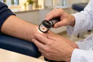
Dermoscopy and comparative photography
Dermoscopy is a non-invasive examination performed with a magnifying instrument that illuminates the skin. It reveals pigmented and vascular structures invisible to the naked eye. In some high-risk patients, standardized photographs or digital mapping can facilitate comparison over time. For complex lesions, confocal microscopy can complement dermoscopy.
Biopsy or excision with histological analysis
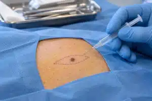
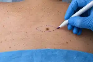
When a lesion appears suspicious, the diagnosis relies on the histopathological analysis of a sample. The physician chooses the biopsy technique based on the size, location, and context. When possible, complete excision of the lesion allows for optimal analysis. The specimen is then examined under a microscope to determine its nature.
If melanoma is diagnosed, subsequent treatment depends in particular on its thickness—measured by the Breslow thickness—and its histological characteristics. Surgical re-excision with appropriate margins may be necessary, according to oncological guidelines.
Can a mole be removed with a laser?
A suspicious pigmented lesion should not be destroyed by laser, electrocoagulation, or any other technique that would prevent its histological analysis. Destroying a lesion without obtaining a biopsy can delay diagnosis and eliminate information essential for treatment.
For a clearly benign lesion, a non-surgical technique may sometimes be considered on a case-by-case basis. This decision should only be made after a sufficiently certain medical diagnosis.
The practical limitations of laser treatment on a pigmented lesion
Three limitations need to be understood. The first is diagnostic: the laser destroys the tissue instead of removing it, making any analysis impossible. If the lesion was a melanoma, the information about its thickness—crucial for subsequent treatment—is permanently lost.
The second is the illusion of healing: a lesion treated with laser visually disappears on the surface, but cells may persist deeper inside. The lesion appears to be treated while it continues to evolve, without any visible warning signs. This is what makes this situation particularly unfavorable.
The third concerns pigmented recurrence: repigmentation after incomplete destruction is difficult to interpret, both clinically and dermoscopy, because the treatment has altered the architecture of
The injury and the usual landmarks no longer apply.
SECURITY MESSAGE
The problem is not the word "laser" itself: it is the destruction of a potentially malignant lesion without the possibility of analysis.
How does the care process unfold, step by step?
Many patients arrive for their appointments without knowing what to expect. Here is the typical course of treatment, from the initial consultation to follow-up.
1. The initial consultation
The examination focuses on the reported lesion, but also on the entire skin surface: a second, previously unnoticed lesion is sometimes discovered during this time. The patient interview clarifies how long the lesion has been present, what has changed, and explores personal and family risk factors.
2. The decision: monitor or collect
Not all atypical lesions are removed. When examination and dermoscopy are reassuring, photographic monitoring may be offered, with follow-up checks at a later date. A biopsy is performed when doubt persists or when the lesion changes—and in this case, it is not delayed.
3. The gesture and the choice of technique
The technique is chosen according to the size, location, and context. Most skin excisions are performed under local anesthesia on an outpatient basis. The incision is oriented along the skin's tension lines to minimize the visibility of the scar, provided that the safety of the excision allows it.
4. Analysis and communication of the result
The specimen is sent to the pathology laboratory. Before leaving the office, the patient must know when the result will be available and how it will be communicated to them. A result is never left without explanation: whether benign or not, it is explained and its implications clarified.
5. Monitoring
Follow-up includes monitoring wound healing and, depending on the histological results, an appropriate skin surveillance program. In cases of melanoma, management continues according to oncological guidelines, with surgical revision if margins require it and a multidisciplinary approach.
WHAT CLINICAL EXPERIENCE TEACHES
Two situations frequently arise in consultations. The first: a patient worried about a perfectly ordinary lesion, in whom examination reveals another lesion elsewhere that truly warranted attention—hence the importance of examining the entire skin and not just what is visible. The second: a lesion treated elsewhere by destruction, without analysis, making it impossible to determine its nature—hence the emphasis on obtaining an analyzable sample.
Does removing a mole leave a scar?
Every skin incision leaves a scar. Its visibility depends on the size of the lesion, its location, the orientation of the incision, the quality of the closure, and the individual healing process of each patient.
In the case of a suspicious lesion, oncological safety remains the priority. The surgeon's role is to perform an appropriate biopsy while optimizing, as much as possible, the placement of the scar, the suturing, and the scar monitoring.
How to monitor your moles?
- Examine your skin in a well-lit room, facing a full-length mirror.
- Observe the face, ears, neck and scalp by parting the hair.
- Check the torso, back and buttocks with a hand mirror or with the help of a relative.
- Examine the arms, armpits, hands and nails.
- Check the legs, the soles of the feet, the spaces between the toes and the nails.
- Photograph a lesion only to document its evolution, without delaying a consultation.
The frequency of self-examination and medical follow-up depends on individual risk. A patient with numerous moles, a history of melanoma, or immunosuppression requires a personalized monitoring program.
When should I consult a doctor in Paris for a suspicious skin lesion?
A consultation is recommended as soon as a lesion changes, bleeds, itches persistently, or is clearly distinct from others. In Paris, Dr. Nathaniel Stroumza-Escoffier treats skin lesions requiring surgical evaluation or excision with histological analysis, adhering to the principles of dermatological surgery and oncological safety.
When the examination requires additional dermatological expertise, specialized mapping or multidisciplinary oncological care, an appropriate referral is organized.
WHEN TO MAKE AN APPOINTMENT
Has your mole changed, bled, or looked different from others? Make an appointment for a medical examination. If in doubt, do not attempt to burn, shave, or treat it yourself.
F.A.Q
How can you tell if a mole is dangerous?
The nature of a mole cannot be confirmed at home. The ABCDE rule, recent changes, and the "ugly duckling" sign can help identify a lesion that warrants examination. Diagnosis is based on a medical examination and, if necessary, histological analysis.
Is a bleeding mole necessarily cancerous?
No. Friction or trauma can cause bleeding. However, spontaneous or repeated bleeding, or bleeding associated with a change in shape, color, or size, warrants prompt medical attention.
Is an itchy mole something to worry about?
An isolated itch may be benign. It becomes a sign of concern when it persists, appears recently, or is accompanied by a visible change, crusting, or bleeding.
Is it normal to get a new mole in adulthood?
The appearance of new moles is common until around age 35–40. A pigmented lesion that is truly new after age 40, or that looks different from others, warrants a medical examination, without necessarily meaning that it is malignant.
Can a melanoma measure less than 6 mm?
Yes. The 6 mm threshold is a guideline, not an absolute rule. A small lesion that changes or stands out from others may be suspicious.
Is it possible to have melanoma without a pre-existing mole?
Yes. The majority of melanomas appear on previously normal skin; only a portion develop from a pre-existing pigmented lesion.
Should all atypical moles be removed?
No. The decision depends on the clinical examination, dermoscopy, the progression of the lesion, and the level of risk. Some lesions are monitored; others require biopsy or excision.
Can a mole be removed with a laser?
A suspicious pigmented lesion should not be destroyed with a laser without histological analysis. For a clearly benign lesion, the technique can be discussed on a case-by-case basis by a physician.
What is the difference between dermoscopy and biopsy?
Dermoscopy allows observation of the lesion without incision. Biopsy removes all or part of the lesion for microscopic analysis, the only method that can definitively establish its histological nature.
Is the excision performed under local anesthesia?
Most small skin excisions are performed under local anesthesia. The specific procedure depends on the size, location, and required intervention.
Is the removal of a suspicious mole covered by insurance?
The excision of a suspicious lesion for diagnostic purposes is a medical procedure, distinct from cosmetic removal. The treatment details are explained during the consultation, and a cost estimate is provided when required by regulations.
How long does it take to get the results of the analysis?
The turnaround time varies depending on the laboratory and the complexity of the analysis. The patient must receive clear information about the expected turnaround time and how the result will be communicated to them.
How often should you have your moles checked?
The frequency depends on the level of risk. People with many moles, atypical moles, a personal or family history of melanoma, or immunosuppression require personalized monitoring.
Can an app or artificial intelligence diagnose melanoma?
An application can draw attention to a lesion, but it should not be used alone to make a diagnosis or delay a consultation. The medical decision is based on clinical examination, dermoscopy, and, if necessary, histological analysis.
WARNING
This article is for informational and educational purposes only. It is not a substitute for individual medical consultation. Nothing on this page can be used to make a diagnosis remotely: the information provided helps identify a lesion that should be examined, not to determine its nature. If you have any doubts, consult a doctor immediately.
Main scientific references
References from peer-reviewed journals and institutional recommendations.
- Melanoma: Diagnosis and Treatment. American Family Physician, 2024. Clinical review on melanoma diagnosis, the ABCDE rule, the ugly duckling sign, dermoscopy and biopsy modalities.
- Atypical Moles. American Family Physician, 2026. Management of atypical nevi, association with the risk of melanoma, recommended excision margins (1 to 3 mm circumferential), role of systematic surveillance.
- American Academy of Dermatology AssociationWhat To Look For: ABCDEs Of Melanoma. Official recommendations on the ABCDE rule and skin self-examination.
- Jütte L., González-Villà S., Quintana J. et al. Integrating generative AI with ABCDE rule analysis for enhanced skin cancer diagnosis. Frontiers in Medicine, 2024. DOI: 10.3389/fmed.2024.1445318.
- Evaluating the Diagnostic Accuracy of Reflectance Confocal Microscopy to Diagnose Skin Cancer. PMC6107731. The role of confocal microscopy as a complement to dermoscopy for difficult lesions.
- Pampena R. et al. A meta-analysis of nevus-associated melanoma. Journal of the American Academy of Dermatology, 2017. Source of the proportion of melanomas occurring on healthy skin.
- National Cancer Institute (INCa)Overview of cancers in France: incidence data for cutaneous melanoma.
- High Authority for Health (HAS)Recommendations regarding cutaneous melanoma.
ABOUT THE AUTHOR
Dr. Nathaniel Stroumza-Escoffier is a plastic surgeon in Paris, qualified in plastic, reconstructive and aesthetic surgery (RPPS 10100674943), former head of clinic-assistant at the Hospitals of Paris (Tenon Hospital, AP-HP) and member of the SOFCPRE.
It manages skin lesions requiring excision and histological analysis, with particular attention paid to oncological safety, quality of closure and scar monitoring.
Office: 11 rue du Faubourg Saint-Honoré, 75008 Paris.
This content was written and then medically reviewed by Dr. Stroumza-Escoffier. Editorial process: literature review using official sources, writing, medical validation, periodic updates.
Article written by the editorial team and approved by Dr. Nathaniel Stroumza-Escoffier — plastic surgeon, qualified in plastic, reconstructive and aesthetic surgery, former assistant clinical head of the Hospitals of Paris (Tenon Hospital, AP-HP) — RPPS 10100674943. Member of the SOFCPRE. Practice: 11 rue du Faubourg Saint-Honoré, 75008 Paris.
Last updated: July 2026.

This article is for informational purposes only and does not replace a medical consultation.